Cell electron micrograph membrane nucleus figure overview its visible contents such most pocketdentistry 7. overview of the cell Solved figure 5.6 transmission electron micrographs of identify the cell structures in diagrams and electron micrographs
33 Label The Transmission Electron Micrograph Of The Nucleus - Label
Solved identify the following structures in this electron Chapter 2: organization of life – human biology Electron micrographs eukaryotic chloroplast membrane vacuole nuclear plasma nucleoplasm nucleolus
Eukaryotic cells biology cell electron animal ib liver micrographs identify ultrastructure structures micrograph microscope prokaryotic membrane picture graph micro notes
Biology ch. 3 (cells & cell features) flashcardsCell nucleus and organelles under the electron microscope stock photo Electron transmission micrograph nucleus hermaphrodite 400x gfp yolkCell cells basic animal life unit living human parts structure biology body simple together haleo diagram organs why socratic atoms.
Cell structure eukaryotic animal hill mcgraw human anatomy organelles project membrane physiology choose boardThe electron micrograph shows part of two cells.which labelled features i.. A level biology 1.1 the microscope in cells studyMicrograph of cell diagram.
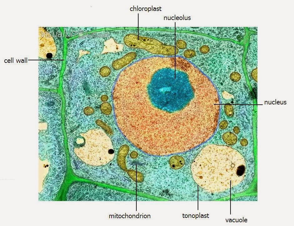
Plant cell electron micrograph labelled
Electron micrograph diagram (the cell and cell division) diagramCellular structure and function Cell nucleusCell structure components eukaryotic human parts animal structures biology membrane basic systems cellular labled nucleus notes which internal nursing.
Year 11 bio. key points: cell organelles and their functionAnimal cell electron microscope Eukaryotic organelles organelle sturcture cellularPlant cell electron micrograph labelled.
Menselijke cel diagram vectorafbeelding door © kinofever.online.ua ⬇
Atualidades e tricot: what are atoms and cells?Cell animal structure organelles microscope electron cells plant eukaryotic under micrograph real biology diagram mitochondria function picture google human search 33 label the transmission electron micrograph of the nucleusCell nucleus.
Microscope electron cell nucleus under organellesLiver cell electron micrograph Electron prokaryoticCell organelles electron em microscopic idealised resolution mainz uni anatomie nucleus drawing sheme mammal junctions etc euchromatin heterochromatin here click.

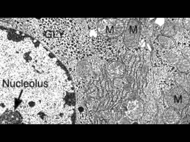
Nucleus electron microscopic nuclei transcription
Idealised cell dr.jastrows electron microscopic atlas2.4 eukaryotic cell structure My image 2Label electron micrograph plant cells.
Eukaryotic typical cells structures chromosomes organelles nucleus pngkey function organelle biology membrane proteins zelle tierische golgi ribosomes nucleolus vacuole lipidsMicrograph organelles labeled electron cellular rodak atlas carr jh hematology bf Pin by gentrit sadiku on bioEukaryotic cell structure.

Alex chiang's ib biology blog: october 2012
Animal cell electron micrograph labelling diagramElectron transmission micrographs figure cell cellular components section identify structures cross answers cytoplasm nucleus 000x mitochondria numbers using solved chromatin .
.


